The nasal bone plays an essential role in shaping the human face, specifically in forming the upper part of the nose. These small, oblong bones are involved in facial structure, protection of the nasal cavity, and attachment for cartilage that shapes the lower nose. Often overlooked due to their size, the nasal bones are structurally and clinically significant. One of the most common anatomical questions regarding the nasal bone is whether it is paired or unpaired. To answer this, we must explore its development, location, function, and its relationship with surrounding structures of the skull.
What Are the Nasal Bones?
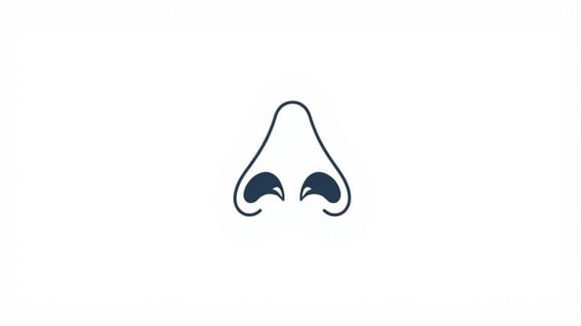
Basic Description
The nasal bones are thin, flat bones located at the bridge of the nose. They are positioned in the midline of the face, just inferior to the glabella (the smooth part of the forehead above the nose) and superior to the nasal cartilage. Their primary job is to form the bony part of the nose and help protect the upper part of the nasal cavity.
Paired Nature of the Nasal Bone
The nasal bone ispaired, meaning that there are two nasal bones one on the left side and one on the right. These two bones are mirror images of each other and meet in the midline at the internasal suture. Together, they form the bridge of the nose. Though the two bones are often fused closely, they are distinct anatomical structures that are symmetrical and develop independently during embryonic growth.
Location and Articulations
Position in the Skull
The nasal bones are part of the facial skeleton and are located between the frontal bone of the forehead and the nasal cartilages of the lower nose. They form the superior portion of the nose’s framework.
Bone Articulations
Each nasal bone articulates with four other structures:
- Superiorly: Articulates with the frontal bone
- Laterally: Articulates with the frontal process of the maxilla
- Medially: Articulates with the opposite nasal bone
- Inferiorly: Connects with the upper lateral nasal cartilage
These articulations contribute to both the stability and mobility of the nasal framework, especially as it relates to facial expression and respiration.
Development of the Nasal Bones
Embryological Origin
The nasal bones develop from the mesenchyme of the frontonasal prominence during embryonic growth. They begin to ossify (turn to bone) in the early fetal stage and continue to grow until adolescence. The bilateral nature of their development is a key indicator of their classification as paired bones.
Fusion and Suture
Even though the nasal bones are paired, they typically appear fused in adult skulls at the midline along the internasal suture. This suture is usually visible on skull X-rays or anatomical models. The fusion may give the illusion of a single bone, but anatomically and developmentally, they remain classified as a pair.
Function of the Nasal Bones
Structural Support
The nasal bones form the upper part of the nasal bridge, giving the nose its shape and supporting the surrounding cartilage and soft tissues. They are crucial for facial symmetry and appearance.
Protection of Nasal Cavity
These bones help protect the upper part of the nasal cavity, shielding it from trauma and environmental exposure. They also serve as a barrier to prevent foreign objects from entering the skull through the nasal passages.
Attachment Point
The nasal bones provide attachment points for nasal cartilages, which form the lower flexible part of the nose. These attachments are essential for functions like breathing and olfaction.
Clinical Relevance
Fractures of the Nasal Bone
Due to their exposed position on the face, nasal bones are the most frequently fractured facial bones. Nasal bone fractures are common in contact sports, accidents, and falls. These fractures can affect both bones simultaneously or just one side.
- Symptoms include swelling, bruising, nasal deformity, and difficulty breathing through the nose.
- Treatment may range from manual realignment to surgical intervention in severe cases.
Nasal Surgery and Cosmetic Procedures
Rhinoplasty, a common plastic surgery procedure, often involves reshaping the nasal bones. Surgeons must have detailed knowledge of the paired structure of these bones to perform safe and effective corrections of the nasal bridge or nasal deviations.
Developmental Anomalies
Occasionally, improper fusion or malformation of the nasal bones can result in congenital conditions such as nasal asymmetry or midline facial defects. These may require surgical correction and are typically diagnosed early in childhood.
Comparison with Unpaired Facial Bones
Paired vs. Unpaired Bones in the Skull
The human skull is composed of both paired and unpaired bones. Examples ofpaired facial bonesinclude:
- Maxilla
- Zygomatic bones
- Palatine bones
- Lacrimal bones
- Nasal bones
Unpaired facial bones include:
- Mandible
- Vomer
The paired nasal bones are symmetrical and articulate with each other, similar to how other paired bones form parts of the face. This bilateral structure is essential for balanced facial features.
Why the Nasal Bone Is Not Unpaired
Though often seen as a single unit in the midline, the nasal bone is not unpaired because it originates from two separate centers of ossification, one on each side of the face. These bones remain anatomically and developmentally distinct, even after fusion at the internasal suture.
Imaging and Anatomical Identification
Radiographic Appearance
In radiographs (X-rays) or CT scans, the nasal bones appear as small rectangular bones at the upper nasal bridge. The internasal suture is usually visible between the two bones. Identifying this suture is important in assessing nasal trauma and planning surgical interventions.
Palpation and Examination
In physical exams, the nasal bones can be palpated easily on the bridge of the nose. Doctors often use this technique to assess alignment, swelling, or tenderness after trauma.
The nasal bone is clearly classified as apaired bonein human anatomy. Although the two nasal bones fuse in the midline to form the bridge of the nose, they originate as separate bones and remain distinct in structure. These paired bones contribute significantly to facial appearance, nasal function, and protection of the nasal cavity. Their anatomical location, function, and vulnerability to trauma make them a subject of interest in medical, surgical, and dental fields. A clear understanding of the paired nature of the nasal bones enhances our appreciation of human craniofacial structure and assists in accurate diagnosis and treatment of nasal conditions.