Paramecium is a fascinating single-celled organism that offers an ideal starting point for students learning about microscopic life. While it might seem complex at first, a simplified diagram of paramecium can make it much easier to understand its basic structure and function. These organisms are classified as protists and live in aquatic environments, where they use tiny hair-like structures to move and feed. Knowing how to interpret or draw an easy diagram of paramecium helps learners visualize biology concepts more effectively and builds a solid foundation in both cell biology and life science.
Understanding the Basics of Paramecium
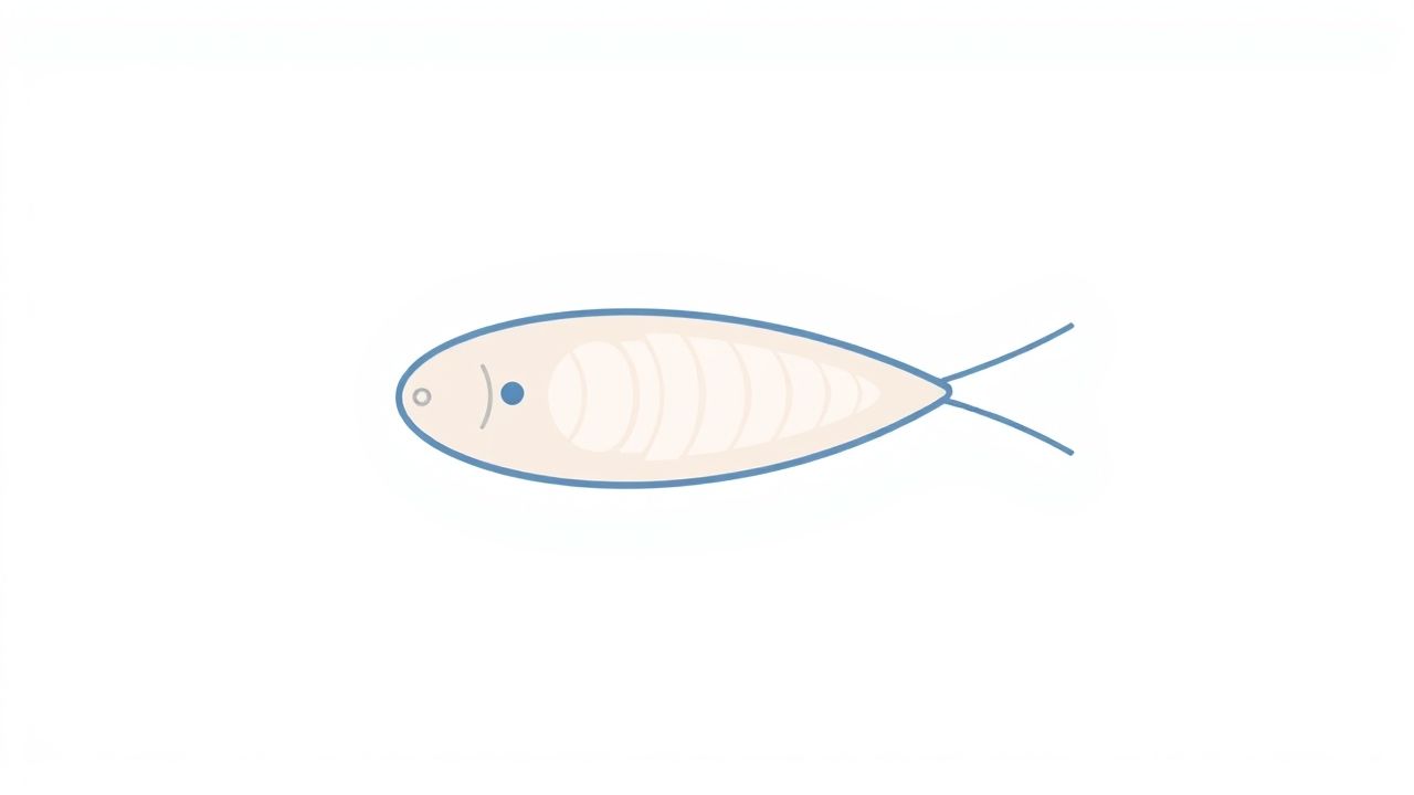
What Is a Paramecium?
Paramecium is a genus of unicellular ciliates, commonly studied as a representative of the protist kingdom. It is known for its slipper-like shape and fast movement through freshwater environments. Paramecia are heterotrophic, meaning they obtain food from their surroundings rather than making it themselves like plants do.
Why Learn with a Diagram?
Diagrams are an excellent learning tool because they simplify complex biological structures. An easy diagram of paramecium can clarify how this organism functions, which parts are responsible for feeding, reproduction, and movement, and how all the internal organelles are arranged. Students of all levels, from middle school to introductory college courses, can benefit from studying or drawing labeled paramecium diagrams.
Key Parts to Include in an Easy Paramecium Diagram
1. Cell Membrane
The outer layer of the paramecium is the cell membrane. It controls the passage of substances in and out of the cell and helps maintain the organism’s shape. In a diagram, this is usually the outermost line surrounding the cell.
2. Cilia
Cilia are tiny hair-like structures that cover the entire surface of the paramecium. They beat rhythmically to propel the organism through water and also help sweep food ptopics into the oral groove. In diagrams, cilia can be drawn as short strokes lining the edge of the organism.
3. Oral Groove
This is an indentation on one side of the cell where food is collected. The cilia push food ptopics into this groove. In the diagram, the oral groove should be marked clearly, often shown as a curved opening near the middle or front of the cell.
4. Gullet and Food Vacuoles
Once food enters the oral groove, it is funneled into the gullet and packaged into food vacuoles. These vacuoles travel through the cell’s cytoplasm, where enzymes digest the food. In the diagram, food vacuoles can be shown as small circular bubbles moving away from the oral groove.
5. Macronucleus and Micronucleus
Paramecia have two types of nuclei: a large macronucleus and a smaller micronucleus. The macronucleus handles everyday cell functions, while the micronucleus is involved in reproduction. In a diagram, the macronucleus should be larger and oval-shaped, while the micronucleus is smaller and usually nearby.
6. Contractile Vacuoles
These star-shaped structures are involved in osmoregulation. They expel excess water to prevent the cell from bursting. Diagrams often place them at either end of the cell, labeled as circular or star-like shapes.
How to Draw an Easy Diagram of Paramecium
Step-by-Step Tips
To draw a simple and accurate diagram of paramecium, follow these easy steps:
- Start by sketching an oval or slipper-like shape to represent the general outline of the organism.
- Add tiny lines or short strokes around the outer edge to show the cilia.
- On one side, draw a curved line to indicate the oral groove.
- Inside the cell, draw two vacuoles and label one as the contractile vacuole and the other as a food vacuole.
- Draw a large oval for the macronucleus and a smaller circle beside it for the micronucleus.
- Include a few circular structures to represent food vacuoles floating in the cytoplasm.
- Label each part clearly for clarity and learning purposes.
Labeling Tips
When labeling, use clean lines and keep labels outside the main body of the diagram to avoid clutter. Make sure all key structures such as cilia, oral groove, nuclei, and vacuoles are identified correctly. Simple labels improve understanding and help during study or exams.
Importance of Diagrams in Science Education
Visual Learning Benefits
Many learners understand better through visual aids than through text alone. A clear and simple paramecium diagram provides an immediate visual connection to the lesson. It reinforces memory by associating names and functions with visible shapes.
Improves Retention and Recall
When students draw or review labeled diagrams regularly, they are more likely to remember the parts and processes of the organism. This is especially useful during quizzes or standardized tests where labeling is required.
Helps in Scientific Communication
Learning to read and draw diagrams fosters scientific communication skills. It trains students to explain biological concepts with clarity, which is a vital skill in both academic and professional settings.
Where to Use Easy Paramecium Diagrams
Educational Applications
Easy diagrams of paramecium are used in textbooks, biology worksheets, and exam preparation materials. Teachers often use these diagrams in classroom presentations or assign students to label them as homework or lab exercises.
Science Projects
Students may include paramecium diagrams in science fair projects to explain unicellular life or demonstrate microscope skills. A well-drawn diagram can help explain the role of protists in ecosystems and their similarities with more complex organisms.
Online Learning Platforms
As online education continues to grow, simple paramecium diagrams are often featured in digital courses and e-learning modules. These visuals enhance learning and are easier to understand on screens when simplified and labeled properly.
Learning about the paramecium becomes much easier with a simplified, easy-to-follow diagram. By understanding the main parts such as the cilia, oral groove, vacuoles, and nuclei, students can appreciate how this tiny organism lives, moves, and feeds. Drawing or reviewing these diagrams reinforces essential biological knowledge and prepares learners for more advanced topics in cell biology. Whether in the classroom, at home, or online, diagrams are a powerful tool for making life science accessible and engaging.