The plantar fascia is a thick, fibrous connective tissue located on the bottom of the foot, playing a key role in supporting the arch and facilitating proper foot mechanics during walking, running, and standing. Its origin and insertion points are vital to understanding both its function and its involvement in common conditions such as plantar fasciitis. This strong band of tissue serves as a bridge between the heel bone and the toes, and is essential for absorbing shock and maintaining the structural integrity of the foot. By studying the origin and insertion of the plantar fascia, we gain deeper insight into how the foot operates in everyday motion and what happens when injuries occur.
Overview of the Plantar Fascia
What Is the Plantar Fascia?
The plantar fascia, also known as the plantar aponeurosis, is not a muscle or tendon, but rather a ligament-like sheet of dense connective tissue. It runs along the bottom of the foot and is critical in arch support. Despite its name, the plantar fascia is composed primarily of collagen fibers and functions similarly to a strong elastic band that stores and releases energy with each step.
Importance in Foot Mechanics
This structure helps stabilize the medial longitudinal arch and supports the foot during the stance phase of gait. It also acts as a lever for propulsion and shock absorber, ensuring smooth movement from heel strike to toe-off.
Origin of the Plantar Fascia
Medial Calcaneal Tubercle
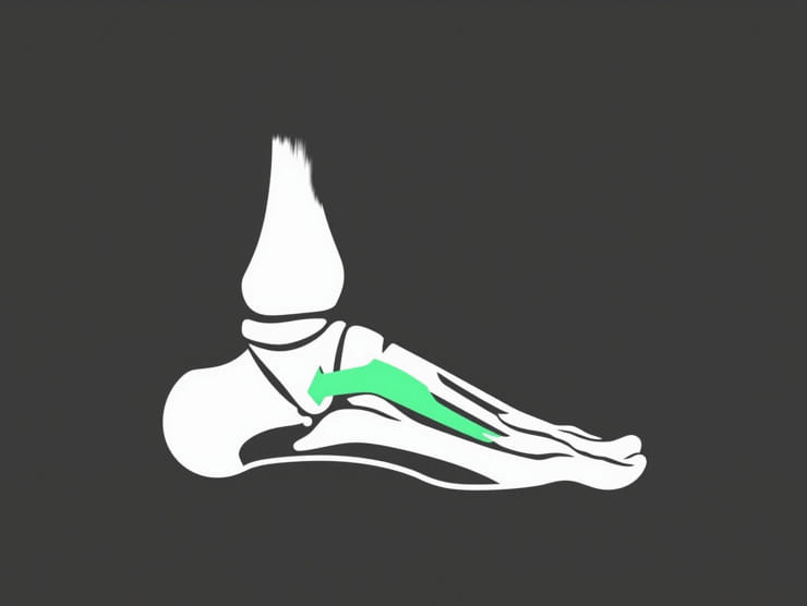
The origin of the plantar fascia is firmly rooted at the medial process of the calcaneal tuberosity, located on the underside of the heel bone. This specific site provides a strong foundation for the fascia to withstand the high tension and forces generated during walking and running.
Why the Origin Matters
The origin point is a common site of inflammation and micro-tears, especially in individuals who suffer fromplantar fasciitis. This repetitive stress condition often manifests as heel pain and is usually due to excessive tension at the calcaneal attachment of the fascia.
Insertion of the Plantar Fascia
Fibrous Slips into the Toes
From its origin at the calcaneus, the plantar fascia extends forward and splits into five distinct bands. Each band inserts into the bases of the proximal phalanges of the toes via the fibrous digital sheaths. These insertions are reinforced by connections with the flexor sheaths and the transverse metatarsal ligaments.
Insertion Sites Summary
- Medial band: inserts into the first toe region (hallux)
- Central band: the thickest, connects to the second, third, and fourth toes
- Lateral band: inserts near the fifth toe
Functional Importance of the Insertion
The insertion of the plantar fascia into the toes is essential for transmitting tension from the heel to the forefoot during gait. When the toes dorsiflex, especially during toe-off, the plantar fascia tightens, increasing the arch height through thewindlass mechanism. This tightness stabilizes the foot, making it a rigid lever for efficient push-off.
Three Distinct Bands of the Plantar Fascia
Central Band
The central band is the thickest and most clinically significant portion. It extends from the calcaneal tuberosity, fans out across the sole, and contributes to the digital fibrous sheaths. It bears most of the weight and tension during standing and movement.
Medial Band
Thinner and less prominent, the medial band runs from the medial calcaneus toward the area near the great toe. While less commonly injured, it plays a supportive role in maintaining the medial arch.
Lateral Band
The lateral portion of the plantar fascia extends toward the lateral aspect of the foot, contributing to lateral arch support and stabilizing the fifth metatarsal.
Relationship with Surrounding Structures
Adjacent Muscles
Though not directly attached to muscles, the plantar fascia works closely with several intrinsic foot muscles including:
- Flexor digitorum brevis
- Abductor hallucis
- Quadratus plantae
These muscles help reduce stress on the plantar fascia during movement.
Neurovascular Structures
The fascia lies superficial to many important structures, such as the lateral and medial plantar nerves and vessels. Understanding this proximity is important in surgical interventions and managing nerve entrapment syndromes.
Plantar Fascia in Gait and Movement
Windlass Mechanism
During walking, the toes extend at the end of the stance phase. This action winds the plantar fascia around the metatarsal heads, tightening it and elevating the arch. This process stores mechanical energy and converts the foot into a rigid lever, essential for efficient propulsion.
Arch Support
The plantar fascia acts like a tie-rod that spans from the calcaneus to the forefoot. As weight is placed on the foot, it helps absorb and redistribute the load while preventing over-flattening of the arch.
Common Injuries and Conditions
Plantar Fasciitis
The most common condition related to the plantar fascia, plantar fasciitis is characterized by inflammation at the origin on the medial calcaneal tubercle. Risk factors include obesity, improper footwear, excessive activity, and tight calf muscles.
Fascial Tears
In rare cases, the fascia can partially or fully tear, either due to trauma or overuse. Symptoms include sharp pain, swelling, and an inability to bear weight on the foot.
Heel Spurs
Chronic stress at the fascia’s origin may lead to bony growths known as calcaneal spurs. These can coexist with plantar fasciitis but are not always the source of pain.
Treatment and Rehabilitation
Stretching and Strengthening
- Calf stretches to reduce strain on the fascia
- Toe curls and towel scrunches to strengthen intrinsic muscles
- Rolling the foot over a frozen bottle to reduce inflammation
Orthotics and Footwear
Supportive shoes and custom orthotic inserts help offload pressure from the heel and distribute forces more evenly across the plantar fascia.
Advanced Interventions
- Physical therapy modalities like ultrasound and manual therapy
- Corticosteroid injections in resistant cases
- Plantar fascia release surgery for chronic, non-responsive pain
The origin and insertion of the plantar fascia play critical roles in foot biomechanics, arch support, and energy transfer during gait. Originating at the medial calcaneal tubercle and inserting into the toes, this connective tissue structure is essential for daily movements and athletic performance. When functioning properly, the plantar fascia helps maintain the integrity and efficiency of the foot. When injured, however, it can significantly impact quality of life. A clear understanding of its anatomical connections, function, and related clinical conditions is fundamental for both healthcare professionals and individuals seeking to maintain healthy, pain-free feet.